 English


Cracked Tooth: When It Hurts to Bite and the X-Ray "Shows Nothing"
A brief pain when biting or releasing that comes and goes may be a crack invisible on X-rays. Signs, diagnosis and treatment options according to the evidence.
It only hurts when you bite certain things, sometimes when you release the bite, and then it disappears for days. You see the dentist, they take X-rays and tell you “nothing shows up.” That puzzling pattern has a name: cracked tooth syndrome. It is one of the hardest causes of dental pain to diagnose, and understanding it can save you months of uncertainty.
Picture this hypothetical scene: Richard is 47 and a big popcorn fan. For a few weeks now, every time he bites something hard on the left side he feels a brief, intense, electric jolt. Oddly, the pain shows up more when he lets go than when he clenches. Cold water bothers him a little too. He already had a checkup, an X-ray was taken, and no decay or infection was found. Richard is starting to think he is imagining it.
He is not imagining it. What he describes is a classic picture that dentistry has recognized for decades.
A name more than half a century old
The term “cracked tooth syndrome” became widely known in 1964, when patients were described with pain on chewing and no visible cause on routine examination (Cameron, 1964). Since then it has been understood that a very fine crack, not always visible to the naked eye or on an X-ray, can run through enamel and dentin and cause intermittent discomfort.
Why does it hurt in such a particular way? One accepted explanation is that when you bite, the fragments on either side of the crack separate slightly and move the fluid inside the dentinal tubules. That movement stimulates the nerves of the pulp. When you release, the fragments snap back together, which explains the well-known “rebound pain” (Banerji et al., 2010a).
Who is more likely to have it?
Reviews on the topic agree on some patterns. Cracks are more common in middle-aged adults and tend to appear in molars, especially lower ones, and in upper premolars (Lubisich et al., 2010). Associated factors include large restorations that weaken the walls of the tooth, clenching or grinding, biting hard objects, and certain cusp shapes that concentrate forces.
An analysis of cases also found a surprising fact: a substantial share of cracked teeth had no restorations or only small ones (Seo et al., 2012). In other words, a “healthy” tooth can crack too if it receives intense, repeated forces.
Signs worth writing down
Because the crack is not always visible, the description of symptoms is one of the most valuable diagnostic tools (Banerji et al., 2010a). These are the most typical clues:
- Sharp, brief pain when biting hard or chewy foods, often at one very specific spot.
- Pain on releasing the bite, more than on clenching.
- Sensitivity to cold, and sometimes to sweets.
- Difficulty pointing out exactly which tooth hurts.
- Discomfort that comes and goes over weeks or months.
A large study from a network of dental practices in the United States examined thousands of teeth with visible cracks and found that not all of them caused symptoms. Certain external features, such as cracks that block the passage of light or that connect to a restoration, were linked to a higher likelihood of pain (Hilton et al., 2017). This helps explain why many people have lines in their enamel without any problem, while others suffer a great deal from nearly invisible cracks.
How to look for a crack you cannot see
Diagnosis usually combines several tests. One of the most useful is asking the patient to bite on a special instrument, cusp by cusp, until the pain is reproduced. Transillumination is also used: the tooth is lit with an intense light, and if there is a crack, the light stops at it. Dyes, magnification, and removing an old restoration to look underneath can also help (Banerji et al., 2010a).
The X-ray is still important, but for another reason: it rarely shows the crack, although it does help rule out other causes of pain and detect bone damage once the crack has progressed (Lubisich et al., 2010). That is why a “clean” X-ray does not mean nothing is there.
Not all cracks are the same
Severity depends on how far the crack extends. A superficial line in the enamel almost never needs treatment. A fractured cusp usually has a good outlook because the fragment is removed and the tooth is restored. A crack that moves toward the center of the tooth can compromise the pulp. And when the crack splits the tooth in two or runs very deep into the root, keeping the tooth may not be possible (Kakka et al., 2022).
That is why timing matters. A crack detected early has many more treatment options than one that kept growing through months of chewing.
Treatment options
The main goal is to hold the fragments together so they stop moving when you bite. When the pulp is healthy, protecting the tooth with a restoration that covers the cusps, such as a crown or an onlay, is often enough. Sometimes a provisional protection is placed first to confirm that the pain goes away before the definitive treatment is done (Banerji et al., 2010b).
Bonded resin restorations have also been studied. In a seven-year follow-up of painful cracked teeth restored with direct composite, most stopped hurting and the restorations kept working, although some needed repairs over time (Opdam et al., 2008).
Not every crack that is found needs immediate treatment. In the same practice network, dentists tended to recommend treatment mainly when there were symptoms, decay or an obvious crack, while many cracks without discomfort were simply monitored (Hilton et al., 2020).
What if the crack reached the nerve?
When the pulp is irreversibly inflamed or infected, a root canal treatment is needed in addition to protection with a crown. The results are encouraging: in one study, about nine out of ten cracked teeth treated with a root canal were successful at one year. Outcomes were worse when there were deep periodontal pockets or several cracks in the same tooth (Krell & Caplan, 2018).
Another study showed high survival of root-canal-treated cracked teeth, lower when the crack extended toward the root (Kang et al., 2016). A systematic review with meta-analysis confirmed those high survival rates and identified pocket depth as one of the most important prognostic factors (Olivieri et al., 2020).
How to protect your teeth
- Avoid chewing ice, olive pits, unpopped popcorn kernels, pens or fingernails.
- If you clench or grind your teeth, tell your dentist; a protective night guard may be recommended.
- Do not put off checking very large or worn restorations.
- If a tooth hurts only when you bite, note which foods trigger it and when the pain occurs; that information is very useful at the appointment.
- While you wait for your visit, chew on the other side to keep the crack from spreading.
Frequently asked questions
Can a crack close on its own? No. Enamel and dentin do not regenerate the way bone does. What can be done is to stabilize the tooth so the crack does not progress.
If the pain stops, does that mean it healed? Not necessarily. The pain can come and go while the crack keeps growing. It is worth completing the assessment.
Are the lines I see on my front teeth dangerous cracks? Often they are superficial enamel lines, very common with age and harmless. Your dentist can tell them apart.
The end of the story
In our hypothetical case, Richard went back with a list of the foods that triggered his pain. His dentist did a cusp-by-cusp bite test and reproduced the exact jolt in a lower molar. Transillumination revealed a fine crack next to an old filling. The pulp responded normally, so a restoration covering the cusps was placed. The pain went away within a few days. Richard learned two things: a clean X-ray does not always tell the whole story, and unpopped popcorn kernels are not as harmless as they look.
If you want a professional to take a look
Learn more about crowns and veneers at The Special Teeth. If you are coming from the United States, our guide to planning your visit to Tijuana helps with the border crossing and transfers. Whenever you are ready, you can request an evaluation.
References
Banerji, S., Mehta, S. B., & Millar, B. J. (2010a). Cracked tooth syndrome. Part 1: Aetiology and diagnosis. British Dental Journal, 208(10), 459–463. https://doi.org/10.1038/sj.bdj.2010.449
Banerji, S., Mehta, S. B., & Millar, B. J. (2010b). Cracked tooth syndrome. Part 2: Restorative options for the management of cracked tooth syndrome. British Dental Journal, 208(11), 503–514. https://doi.org/10.1038/sj.bdj.2010.496
Cameron, C. E. (1964). Cracked-tooth syndrome. The Journal of the American Dental Association, 68(3), 405–411. https://doi.org/10.14219/jada.archive.1964.0108
Hilton, T. J., Funkhouser, E., Ferracane, J. L., Gilbert, G. H., Baltuck, C., Benjamin, P., Louis, D., Mungia, R., & Meyerowitz, C. (2017). Correlation between symptoms and external characteristics of cracked teeth. The Journal of the American Dental Association, 148(4), 246–256.e1. https://doi.org/10.1016/j.adaj.2016.12.023
Hilton, T. J., Funkhouser, E., Ferracane, J. L., Schultz-Robins, M., Gordan, V. V., Bramblett, B. J., Snead, R. M., Manning, W., & Remakel, J. R. (2020). Recommended treatment of cracked teeth: Results from the National Dental Practice-Based Research Network. The Journal of Prosthetic Dentistry, 123(1), 71–78. https://doi.org/10.1016/j.prosdent.2018.12.005
Kakka, A., Gavriil, D., & Whitworth, J. (2022). Treatment of cracked teeth: A comprehensive narrative review. Clinical and Experimental Dental Research, 8(5), 1218–1248. https://doi.org/10.1002/cre2.617
Kang, S. H., Kim, B. S., & Kim, Y. (2016). Cracked teeth: Distribution, characteristics, and survival after root canal treatment. Journal of Endodontics, 42(4), 557–562. https://doi.org/10.1016/j.joen.2016.01.014
Krell, K. V., & Caplan, D. J. (2018). 12-month success of cracked teeth treated with orthograde root canal treatment. Journal of Endodontics, 44(4), 543–548. https://doi.org/10.1016/j.joen.2017.12.025
Lubisich, E. B., Hilton, T. J., & Ferracane, J. (2010). Cracked teeth: A review of the literature. Journal of Esthetic and Restorative Dentistry, 22(3), 158–167. https://doi.org/10.1111/j.1708-8240.2010.00330.x
Olivieri, J. G., Elmsmari, F., Miró, Q., Ruiz, X.-F., Krell, K. V., García-Font, M., & Durán-Sindreu, F. (2020). Outcome and survival of endodontically treated cracked posterior permanent teeth: A systematic review and meta-analysis. Journal of Endodontics, 46(4), 455–463. https://doi.org/10.1016/j.joen.2020.01.006
Opdam, N. J. M., Roeters, J. J. M., Loomans, B. A. C., & Bronkhorst, E. M. (2008). Seven-year clinical evaluation of painful cracked teeth restored with a direct composite restoration. Journal of Endodontics, 34(7), 808–811. https://doi.org/10.1016/j.joen.2008.04.011
Seo, D.-G., Yi, Y.-A., Shin, S.-J., & Park, J.-W. (2012). Analysis of factors associated with cracked teeth. Journal of Endodontics, 38(3), 288–292. https://doi.org/10.1016/j.joen.2011.11.017
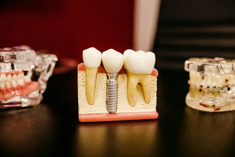
Main image: Yasaman Abedini / Unsplash. Photos are illustrative stock images: they do not show patients, cases or results from The Special Teeth.
Educational information. A professional assessment determines the diagnosis and the appropriate options for each person.